Echocardiogram
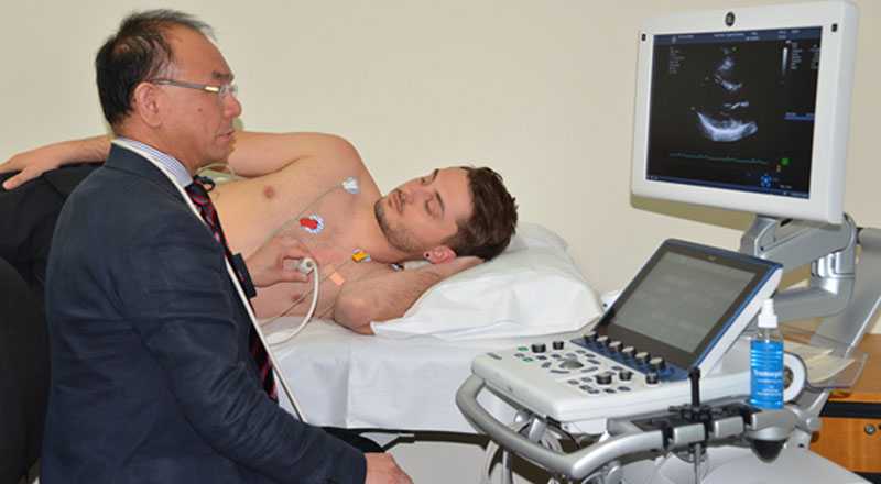
An echocardiogram is an ultrasound of the heart just like any other ultrasound of the body (For example an ultrasound done during pregnancy to monitor the health of a baby). It uses inaudible sound waves which travel through the tissues to create moving images of the heart and its various structures. Its a common test that allows your doctor to see your heart in motion as it beats and pumps blood. It’s a very useful and harmless test to look for any abnormalities in the heart size and function, its structure, chambers, muscle and valves.
Usually a technologist performs the test, a cardiologist reads, interprets and reports. your referring doctor is the right person to discuss the report. The technologist doing the test is not allowed to discuss any part of the study with the patient as the final report may be different depending on the cardiologist’s assessment and interpretation.

Why to do it
There are a range of indications where your doctor may order an echocardiogram, or “echo” (as its commonly called). These include to measure the size of various chambers of the heart, asses overall pumping action of the heart and its various chambers, and alsoe back pressure into the lungs, assess any murmurs and study the valves of the heart – to look for any leaking or obstructing valve conditions, look for any holes in the heart or identify other structural problems like thickness of heart muscle due to hyigh blood pressure, measure pressures in the heart chambers; and a host of other reasons.
Procedure
1. Please make sure you make an appointment for this test.
2. There is no special preparation needed for this test. There is no need to fast.
3. It is a painless non-invasive test. It has no side effects and is completely safe just like the one done during pregnancy.
4. The upper half of your body will need to be exposed for access. You may be provided with a disposable gown for comfort.
5. Its usually done in a dark room.
6. You will be lying on a bed on your left side with left arm under your head and right arm straight.
7. A Technologist will do the ultrasound, sitting on your left side.
8. Three electrodes are usually attached to your body for ECG recording while ultrasound images are being taken
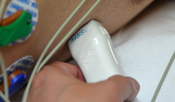
9. A special probe is used which is attached to the echo machine.
10. A small amount of gel is used to lubricate the tip of the probe (it may feel cold).
11. The probe is placed on various areas of the chest and neck, and images are obtained. These are moving images of the heart and also some “Doppler” images which produce audible sound that you can hear during the test.
12. Various positions can be used by the technologist to obtain better images and he/she may ask you to hold you breath at times to record a moving clip.
13. The whole test may take 30 to 40 minutes to complete.
14. After the test the gel is wiped off, ECG electrodes are removed and you will be allowed to dress up and go.
15. No special precautions are needed after the procedure.
16. The report will be finalized by the cardiologist in next 2-3 days and sent to your referring doctor.
17. You should make an appointment with your referring doctor to discuss the results.
Transducer

Getting the best cardiac care
At HeartWest we ensure high standard of our technologists and all reporting cardiologists either hold a Fellowship in Echocardiography or possess DDU (Diploma in Diagnostic Ultrasound) to report Echos to maintain the quality of our cardiac care.
